.png)
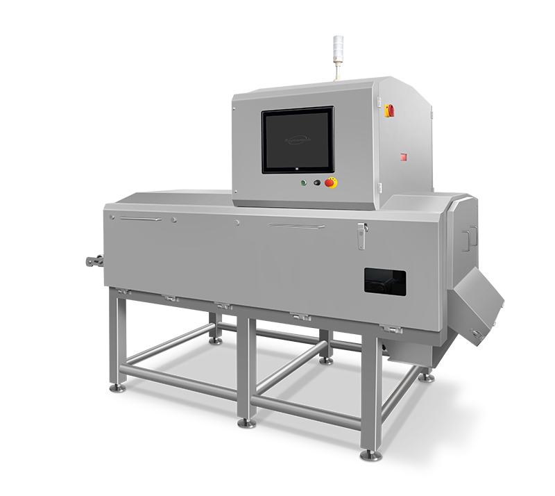
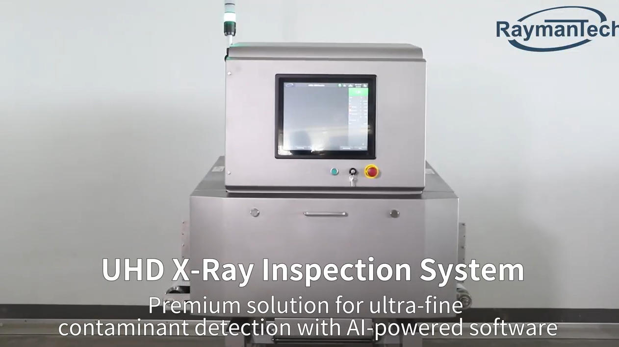
AI + Ultra-HD: The Ultimate AI Optical Sorter for Wet Processed Meat Lines
Optical sorting technology has become indispensabl...
More
Frozen block x-ray methods provide detailed internal views that reveal hidden changes in microstructure.
Over the past decade, X-ray imaging has improved speed and resolution, supporting precise studies in science and industry.
Non-destructive techniques like micro-computed tomography now allow researchers to observe internal features efficiently.
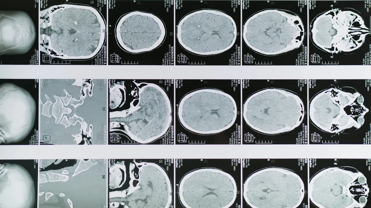
Frozen block x-ray imaging enables researchers to visualize internal transformations that occur during freezing and thawing. This technique leverages high-resolution micro-computed tomography (micro-CT) to produce three-dimensional images of frozen materials. Scientists can observe dynamic processes without altering the sample, thanks to the non-destructive nature of x-ray imaging. Advanced systems, such as 4D micro-CT, allow for the observation of changes over time, providing a deeper understanding of microstructural evolution.
Note: X-ray fluorescence microscopy offers high-resolution imaging while preserving biological samples, unlike traditional methods that often require destructive sample preparation.
The physical principles behind frozen block x-ray imaging rely on the interaction of x-rays with varying densities and compositions within the frozen block. Changes in density and composition can complicate the detection of contaminants, as dense regions may obscure small fragments or low-density materials. Modern inspection systems address these challenges by using multi-energy detection and adaptive algorithms to differentiate between product signals and contaminants.
Researchers commonly observe several types of microstructural changes in frozen blocks using x-ray imaging:
Formation of extremely large pores in samples without annealing, which differ in size and shape from smaller pores.
Reduction in the total number of pores as the amplitude and duration of freeze-thaw cycles increase.
Microstructure of freeze-dried matrices that do not perfectly replicate ice microstructures, indicating significant changes due to water removal.
Micro-CT and image analysis have proven effective for analyzing these microstructural features in frozen materials. However, x-ray imaging does have limitations. Resolution constraints, the need for vacuum pretreatment, and the risk of altering sample morphology during preparation can affect the accuracy of results.
Microstructural changes in frozen blocks play a critical role in determining the quality and properties of materials. In food preservation and material science, the freezing method directly influences ice crystal formation and the resulting microstructure. The following table summarizes the impact of different freezing methods:
| Freezing Method | Ice Crystal Formation | Impact on Microstructure |
|---|---|---|
| Slow Freezing | Large extracellular ice crystals | Structural damage, poor texture, nutrient loss |
| Fast Freezing | Small intracellular and extracellular ice crystals | Minimal damage, better preservation of quality |
Microstructural changes also affect the mechanical properties of frozen blocks. For example:
Increased porosity and permeability in rocks after multiple freeze-thaw cycles lead to deterioration of dynamic strength and elastic modulus.
Porosity can rise to 16.25%, and permeability can increase by 128% after 40 freeze-thaw cycles, indicating significant degradation.
Dynamic strength and elastic modulus decrease as microstructural damage increases.
Frozen block x-ray imaging provides valuable insights into these transformations, supporting improvements in food quality, safety, and material durability. Researchers and industry professionals rely on this technology to optimize freezing processes and enhance product performance.
X-rays interact with frozen materials through absorption and scattering, which depend on the physical and chemical properties of the sample. Temperature and phase changes in frozen blocks, such as the transition from liquid to ice, cause significant shifts in specific volume. For example, the Lα-to-Pβ′ transition of DPPC results in a decrease in specific volume by about 4%, while the water-to-ice transformation increases volume by nearly 10%. These changes alter how X-rays pass through the material, affecting both absorption and scattering. High-pressure phases that develop during freezing can also influence the stability of biopharmaceuticals, which impacts their X-ray characteristics.
Several mechanisms govern X-ray interaction with ice and other frozen substances:
The quasi-liquid layer on ice crystal surfaces plays a key role in how antifreeze proteins (AFPs) interact with ice, as AFPs compete with growing crystals for water molecules.
Hydrophobic interactions and hydrogen bonding influence the growth of ice crystals.
Non-AFP proteins accumulate near ice in the liquid portion, where they experience destabilizing stresses from environmental changes.
Impurities in frozen materials, such as solutes or contrast agents, tend to aggregate during freezing. This aggregation enhances image contrast, making internal structures more visible during frozen block x-ray imaging.
X-ray imaging preserves the natural structure of biological samples at cryogenic temperatures. This advantage prevents dehydration, which often causes artifacts in other imaging methods. As a result, researchers can observe frozen samples in their true state. Recent advances in synchrotron X-ray radiography allow for real-time, in-situ observations of dynamic processes. The high photon flux of synchrotron X-rays shortens imaging times, making the technique efficient for studying frozen samples. Direct, real-time imaging eliminates the need for sample dehydration and provides accurate views of biological structures. These features make frozen block x-ray a superior choice over optical or thermal imaging for frozen materials.
Radiography provides a straightforward method for visualizing the internal structure of frozen blocks in two dimensions. This technique uses X-rays to create images that reveal differences in density and composition. The resolution of radiography plays a crucial role in detecting microstructural changes. Higher contrast-to-noise ratio (CNR) values indicate better sensitivity to small features. The following table compares the sensitivity of different radiography types:
| Radiography Type | CNR Value | Sensitivity to Microstructure |
|---|---|---|
| X-ray Dark-field | 2.5 | High |
| Conventional X-ray | 0.2 | Low |
Researchers often select radiography for quick assessments or when they need to screen large numbers of samples efficiently.
Micro-CT enables detailed three-dimensional analysis of frozen block microstructures. This method allows scientists to visualize and analyze intricate features, such as the sublimation front and pore networks, without physically altering the sample. Specialized equipment maintains subzero temperatures during scanning, which preserves the true structure of frozen samples. Most scanners achieve voxel sizes of 10 μm or less, providing high-resolution images. These capabilities make micro-CT ideal for studying dynamic changes during freeze-drying and for obtaining accurate measurements of internal features.
Dark-field imaging offers enhanced contrast by detecting strong X-ray scattering from ice crystals. This approach reveals unique details that conventional X-ray techniques may miss. The table below highlights the differences:
| Aspect | Dark-Field Imaging | Conventional X-ray Techniques |
|---|---|---|
| Contrast Mechanism | Utilizes strong X-ray scattering from ice crystals | Relies on changes in density leading to low contrast |
| Detection of Freezing | Enhanced detection of freezing processes | Limited to hypoattenuating regions due to density changes |
| Application | Effective for distinguishing frozen from unfrozen tissue | Primarily used for general imaging without specific freezing detection |
Frozen block x-ray methods that incorporate dark-field imaging provide researchers with superior tools for distinguishing subtle microstructural changes.
Researchers have observed distinct patterns in ice crystal formation using frozen block x-ray imaging. Small ice crystals, which possess high surface free energy, tend to melt or sublimate. Larger crystals grow as a result, a process known as Ostwald ripening. This phenomenon becomes especially important in frozen foods, where temperature fluctuations during storage accelerate crystal growth. During thermal cycling, the size of ice crystals increases in a linear fashion, while the number of crystals stabilizes. These findings highlight the importance of controlling temperature to maintain product quality.
Frozen block x-ray studies reveal significant changes in material properties during freeze-thaw cycles. The following tables summarize these transformations:
| Property | Change | Description |
|---|---|---|
| Indentation Modulus | Decrease | Gradual reduction, up to 38% after 1500 cycles |
| Hardness | Decrease | Follows the same trend as indentation modulus |
| ITZ Thickness | Increase | Rapid growth from 25 to 50 μm after 1500 cycles |
| Pore Structure | Change | Micropores merge to form larger pores in mortar and ITZ |
| Property | Change | Description |
|---|---|---|
| Particle Arrangement | Complex | Morphology becomes more intricate |
| Inter-particle Connections | Loose | Connections between particles weaken |
| Pore Morphology | Irregular | Pore shapes become more irregular |
| Surface Porosity | Increase | Overall porosity rises |
| Pore Size Distribution | Change | More large and medium pores appear |
Studies use advanced techniques to measure microstructural changes in frozen blocks. Researchers have found that immersion freezing preserves microstructure integrity, which is crucial for food quality. Different freezing conditions produce varying ice crystal sizes and distributions. Analytical methods such as SEM, MIP, and micro-CT allow scientists to quantify pore characteristics and micromechanical properties. These measurements help explain how microstructure changes lead to complex mechanical behavior in frozen soils and other materials.
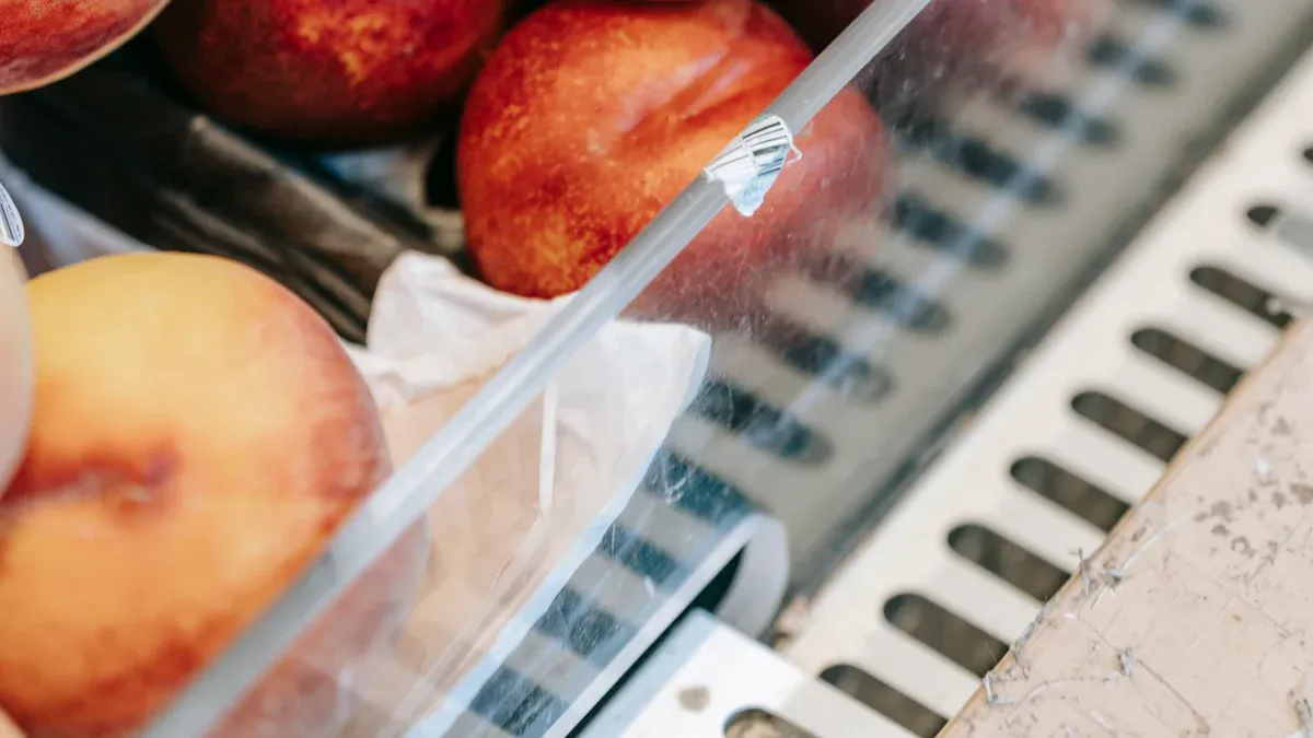
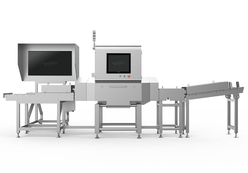
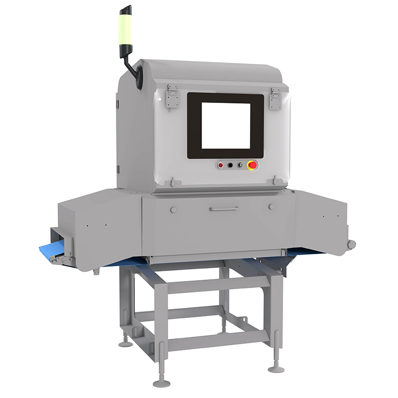
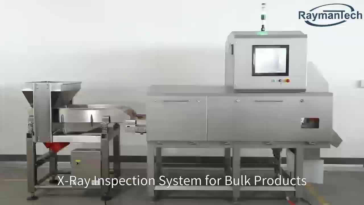
Frozen block x-ray technology has transformed food safety and quality assurance. Food manufacturers use this method to detect contaminants that range from metal fragments to non-metallic materials. The technology also verifies product mass and packaging integrity, which supports consistent quality. Companies can meet international food safety standards, such as HACCP, BRC, and IFS, by implementing these advanced inspection systems. The following table highlights the main capabilities that drive improvements in food quality and safety:
| Capability | Description |
|---|---|
| Contaminant Detection | Detects both metallic and non-metallic contaminants, ensuring comprehensive safety checks. |
| Compliance with Regulations | Helps manufacturers meet international food safety standards like HACCP, BRC, and IFS. |
| Quality Control | Verifies product mass, checks for underfilled or overfilled packages, and ensures packaging integrity. |
| Efficiency | Streamlines production by reducing the need for multiple inspection devices, saving space and time. |
| Versatility | Inspects through various packaging types, including foil and glass, which are challenging for other methods. |
Researchers have made significant progress in material science by using frozen block x-ray imaging. X-ray tomography now maps both crystalline and amorphous phases in frozen biomaterials. This capability helps scientists understand how freezing affects the structure and function of materials. In cryopreservation, the use of inductive heating of magnetic nanoparticles has improved cell and tissue revitalization. This advance may change tissue and organ transplantation by removing time limits between harvesting and implantation. These breakthroughs support the development of safer and more effective preservation techniques.
X-ray tomography maps crystalline and amorphous phases in frozen biomaterials.
Inductive heating of magnetic nanoparticles improves cell and tissue revitalization.
These advances may change approaches to tissue and organ transplants.
Frozen block x-ray research has influenced many industries beyond food and materials. Pharmaceutical companies use x-ray imaging to detect particulates in lyophilized products, which improves quality control. The food industry benefits from better process monitoring, as the technology can find contaminants like wood and insects. Automated detection systems, powered by x-ray imaging, streamline inspection processes and increase efficiency in both fields.
X-ray imaging enhances quality control in pharmaceuticals.
The technology improves process monitoring in food production.
Automated systems support faster and more reliable inspections.
Frozen block x-ray imaging reveals microstructural changes that drive innovation in food technology, material science, and preservation.
Researchers use these insights to improve product quality and safety.
Ongoing studies promise new applications and greater benefits.
Note: This technology continues to shape scientific and industrial progress.
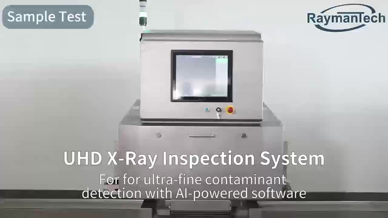
X-ray imaging provides non-destructive, high-resolution views of internal structures. Researchers can detect microstructural changes and contaminants without altering or damaging the sample.
Micro-CT generates three-dimensional images with fine detail. Conventional radiography produces two-dimensional images, which may miss subtle internal features in frozen samples.
X-ray imaging detects most metallic and non-metallic contaminants. Very low-density materials, such as some plastics, may remain challenging to identify.

We provide you with comprehensive foreign trade solutions to help enterprises achieve global development
Automatic recognition and rejection, fish bone ins...
Uneven and overlapped product detection, thin & lo...
.png)
Glass-in-glass / Metal-in-metal inspection, small ...
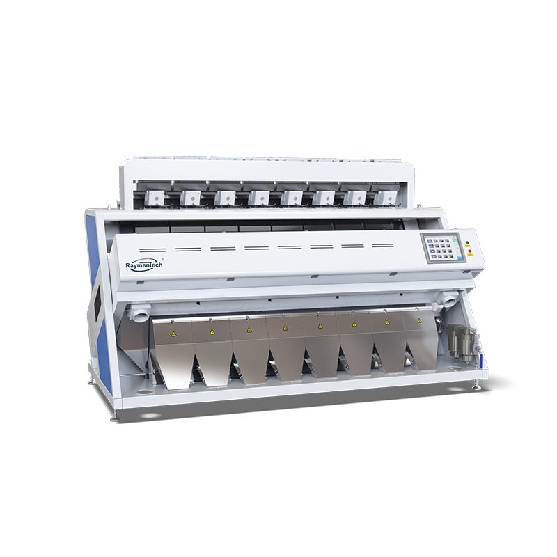
Recommedation: Rice, wheat, corn, grain, pulses, s...
Select the most popular foreign trade service products to meet your diverse needs
Select the most popular foreign trade service products to meet your diverse needs


User Comments
Service Experience Sharing from Real Customers
Michael Chen
Laboratory ManagerThe frozen block x-ray system has revolutionized our sample screening process. The image clarity on cryo-preserved tissues is exceptional, allowing for precise localization before sectioning. A game-changer for our lab.
Sarah Johnson
Quality Control InspectorHighly efficient for non-destructive inspection of frozen food blocks. It helps us detect foreign objects and density inconsistencies quickly, ensuring product safety without thawing. The interface is user-friendly.
David Rodriguez
Forensic PathologistThis imaging device provides invaluable insights in forensic examinations. We can now x-ray frozen biological specimens in situ, preserving evidence integrity and revealing details missed by traditional methods. Outstanding tool.
Emily Watson
Pharmaceutical AnalystExcellent for analyzing the internal structure and homogeneity of frozen drug formulation blocks during R&D. The high-resolution scans save us significant time and materials in the development cycle. Very reliable.