.png)
Meat Packaging Revolution: Mastering Detection Challenges with Next-Gen X-ray and Vision Systems
This article delves into the persistent pain point...
More
Knee cartilage problems affect millions worldwide. The cartilage detection system uses radiomics and machine learning to streamline knee assessments. Clinicians see improved accuracy and efficiency, while patients experience greater comfort. The growing concern is clear.
| Metric | Value |
|---|---|
| Global prevalence of knee OA | 16.0% (95% CI, 14.3%-17.8%) |
| Prevalence in individuals aged 40+ | 22.9% (95% CI, 19.8%-26.1%) |
| Individuals with knee OA (40+) | ~654.1 million (2020) |
Manual cartilage assessment presents several challenges for clinicians and patients. Traditional radiographic methods only offer indirect visualization of cartilage, which limits diagnostic accuracy. Operators often spend significant time segmenting cartilage on MR images. This process requires extensive training and can lead to inconsistent results. Poor reproducibility and low sensitivity to change further complicate manual assessments.
Traditional radiographic measurement provides only indirect visualization of cartilage.
Manual segmentation of cartilage on MR images is time-consuming and burdensome.
Extensive training is required for operators using cartilage segmentation software.
Traditional methods show poor reproducibility and sensitivity to change.
Comparative studies highlight the advantages of automated systems over manual methods. The table below summarizes key findings from recent research:
| Study | Method | Findings |
|---|---|---|
| Bowes et al. | Automated vs. manual | Strong correlations; greater sensitivity to change in automated measures. |
| Wirth et al. | DL-based T2 vs. manual | Similar sensitivity; greater accuracy with advanced neural networks. |
| Eckstein et al. | DL-based vs. manual | Comparable sensitivity; no significant differences in discriminative power. |
| Panfilov et al. | DL-based vs. manual | Strong correlations; lower odds ratios for DL-based technique. |
Current imaging techniques often lack the sensitivity needed to detect early cartilage degeneration. Clinicians recognize the value of a multimodal imaging approach, which combines MRI and CT to provide comprehensive information about both cartilage and bone. The cartilage detection system addresses these gaps by offering faster, more accurate, and reproducible assessments. This innovation supports better clinical decisions and improves patient outcomes.
Radiomics technology transforms medical imaging by extracting quantitative data from standard scans. This approach goes beyond what the human eye can see, converting images into high-dimensional data that reveal subtle patterns in tissue structure. In the context of knee health, radiomics enables clinicians to detect early cartilage changes that traditional imaging might miss.
Radiomics plays a pivotal role in the cartilage detection system. It leverages advanced algorithms to analyze MRI and CT images, identifying features that indicate cartilage health or degeneration. Recent studies have demonstrated the effectiveness of radiomics in early identification and progression stratification of knee osteoarthritis. For example, a two-stage radiomics model can predict structural progression by focusing on features from both cartilage and subchondral bone. Another study introduced a novel radiomic analysis approach, using both CT and MRI, to assess cartilage degradation and establish reliable biomarkers for early osteoarthritis detection.
| Study Title | Focus | Findings |
|---|---|---|
| MRI-based radiomics framework for early identification and progression stratification in knee osteoarthritis | Knee osteoarthritis | Developed a two-stage radiomics model to predict structural progression, emphasizing the role of radiomic features from cartilage and subchondral bone in early detection. |
| Innovative Diagnostic Approaches for Predicting Knee Cartilage Degeneration in Osteoarthritis Patients | Cartilage degradation in knee OA | Explored a novel radiomic analysis approach using CT and MRI to assess cartilage degradation, aiming to establish reliable biomarkers for early detection of OA-related changes. |
Peer-reviewed research supports the use of radiomics in cartilage detection systems. These studies highlight the ability of radiomics to enhance early detection and provide insights that traditional imaging cannot offer.
| Study Title | Summary |
|---|---|
| An MRI-based radiomics framework for early identification and progression stratification in knee osteoarthritis | This study presents a two-stage MRI-based radiomics framework that enhances early detection and progression stratification of knee osteoarthritis, supporting clinical screening and intervention. |
| The use of radiomic analysis of magnetic resonance imaging findings in predicting features of early osteoarthritis of the knee | This study explores the potential of radiomics to enhance diagnostic capabilities for early osteoarthritis, providing insights that are not visible to traditional imaging methods. |
| Quality appraisal of radiomics-based studies on chondrosarcoma using METhodological RadiomICs Score (METRICS) and Radiomics Qual... | This study reviews the application of radiomics in chondrosarcoma, emphasizing its potential for classification and prognostication, while addressing challenges in clinical translation due to low scientific quality. |
Note: Radiomics captures information that often remains invisible in conventional imaging, making it a valuable tool for early intervention and personalized treatment planning.
The cartilage detection system relies on radiomics to extract a wide range of features from knee images. These features provide a comprehensive view of cartilage health, supporting more accurate and granular assessments.
| Feature Type | Description |
|---|---|
| First-order features | Basic statistical measures of the intensity values in the image. |
| Texture features | Measures that capture the spatial arrangement of pixel intensities, providing information on patterns. |
| Wavelet transform | A method used to extract features that capture both frequency and location information. |
| Laplacian of Gaussian | A technique used to enhance edges and extract features related to the structure of the cartilage. |
| Selected features | Nineteen features for cartilage and thirteen for subchondral bone were identified for modeling. |
Radiomics improves the granularity and accuracy of cartilage detection in several ways:
It captures texture, intensity, and shape characteristics from imaging data, enhancing the detail of cartilage assessment.
Features such as the gray-level co-occurrence matrix (GLCM) quantify collagen disorganization, offering insights into degenerative changes.
Histogram metrics correlate with proteoglycan content, improving the accuracy of cartilage health assessments compared to traditional imaging.
The integration of these detailed features allows the cartilage detection system to deliver precise, reproducible results. Clinicians can identify early signs of degeneration, monitor disease progression, and tailor treatment strategies with greater confidence.
Machine learning algorithms play a crucial role in the cartilage detection system. These algorithms process large volumes of radiomic data extracted from knee MRI images. They identify patterns and features that indicate cartilage health or degeneration. Researchers have developed models that use up to 1,049 radiomic features, including first-order statistics, shape, texture, and higher-order statistics. These features provide a comprehensive view of the knee joint.
The process begins with feature extraction. Algorithms analyze MRI images and derive quantitative data. They then select the most relevant features for distinguishing between healthy and diseased cartilage. Models such as logistic regression, K-nearest neighbor, and support vector machine (SVM) use these features to classify knee conditions. Tenfold cross-validation helps identify the best features, while randomization manages training time and computational demands.
The table below highlights machine learning algorithms commonly used in cartilage detection:
| Algorithm | Application in Cartilage Detection |
|---|---|
| XGBoost | Used in studies for knee osteoarthritis and cartilage degeneration |
| CatBoost | Effective in analyzing radiomic data for cartilage detection |
| SVM | Utilized for evaluating cartilage and subchondral bone morphology |
These algorithms enable the cartilage detection system to deliver accurate and reliable results. They support clinicians by providing objective assessments based on complex image data.
Automation transforms the workflow of cartilage assessment. The cartilage detection system uses artificial intelligence to segment and quantify cartilage structures automatically. This approach reduces the need for manual annotation, which often takes significant time and requires specialized training.
AI-driven methods can analyze imaging data to detect early signs of osteoarthritis with high accuracy. They support timely and informed treatment decisions. Automation also improves the reliability of cartilage detection by minimizing human error and variability.
Published results demonstrate the effectiveness of these automated systems:
Automatic segmentation of cartilage in the OAI dataset.
Quantification of cartilage thickness and T2 changes over time.
Development of methods for subregional morphological assessment to detect focal cartilage changes.
The following table summarizes the accuracy of advanced machine learning models in cartilage detection:
| Model | DSC (Femoral Cartilage) | DSC (Tibial Cartilage) | Overall DSC | Overall IoU |
|---|---|---|---|---|
| TransUNet | 0.823 | 0.803 | 0.813 | 0.692 |
Another model, based on gender, predicts cartilage volume loss with high accuracy for both men and women:
| Model | R (Testing Stage) | R (Validation) | Notes |
|---|---|---|---|
| Gender-based model | ≥ 0.79 | ≥ 0.78 | Predicts cartilage volume loss with good accuracy for both genders. |
AI technologies enhance the segmentation and quantification of cartilage structures, leading to improved diagnostic accuracy and faster analysis.
The cartilage detection system streamlines the diagnostic process. Clinicians receive rapid, reproducible results. Patients benefit from early diagnosis and more effective treatment plans.
Clinicians follow a streamlined workflow when using the cartilage detection system. This process leverages advanced imaging and machine learning tools to deliver precise results:
Apply the logarithm-processed Navia color-map to visualize T2 maps.
Process imaging data using MATLAB R2022a.
Develop a Python-based pipeline with the Deep Open-Source Medical Analysis framework.
Segment cartilage into binary masks using the DL Knee Segmentation model.
Reformat CartiGram and adjust laterality for consistency.
Align the CUBE volume and mask with CartiGram.
Perform 3D rotation, translation, and isotropic scaling of the CUBE volume.
Use the Mattes mutual information metric for multimodal registration.
Employ Elastix and Transformix libraries for registration and transformation.
Conduct mono-exponential T2 fitting within each masked compartment.
Project T2 maps onto 2D axial and coronal views with the Navia color-map.
Analyze parameters by dividing subregions for femoral and patellar cartilage.
Save cropped T2 maps in DICOM format for review.
The following table highlights how the system simplifies clinical practice:
| Evidence Description | Details |
|---|---|
| Methodology | The system uses advanced methods like Ensemble Empirical Mode Decomposition and CNNs for cartilage damage detection. |
| Accuracy | The SVM classifier achieves an average accuracy of 87% with an AUC of 0.91, supporting reliable diagnosis. |
| Time Efficiency | VAG signal measurements occur one day before surgery, reducing the time between diagnosis and treatment. |
Patients benefit from a rapid and comfortable experience. The system classifies all image patches for a knee joint in about two seconds. Evaluation of the entire joint takes only a few seconds, making it an efficient screening tool for cartilage lesions. This speed reduces waiting times and supports early intervention.
Patients experience a streamlined process, with minimal discomfort and quick results, enhancing overall satisfaction with knee health assessments.
Clinicians value efficiency in diagnostic workflows. The cartilage detection system streamlines cartilage assessment by reducing scan and analysis times. The MIXTURE sequence platform enables simultaneous morphologic and quantitative evaluation, which supports faster decision-making in clinical settings. Studies show that this approach maintains diagnostic accuracy while saving time during MRI procedures.
| Evidence Description | Implication on Efficiency and Time Savings |
|---|---|
| The MIXTURE sequence platform allows for time-efficient simultaneous assessment. | Suggests increased efficiency in cartilage detection processes. |
| Morphologic and quantitative assessments are performed in clinically feasible time. | Indicates potential time savings in MRI scanning procedures. |
| Comparable results between MIXTURE and reference sequences for cartilage defects. | Supports diagnostic utility without compromising time efficiency. |
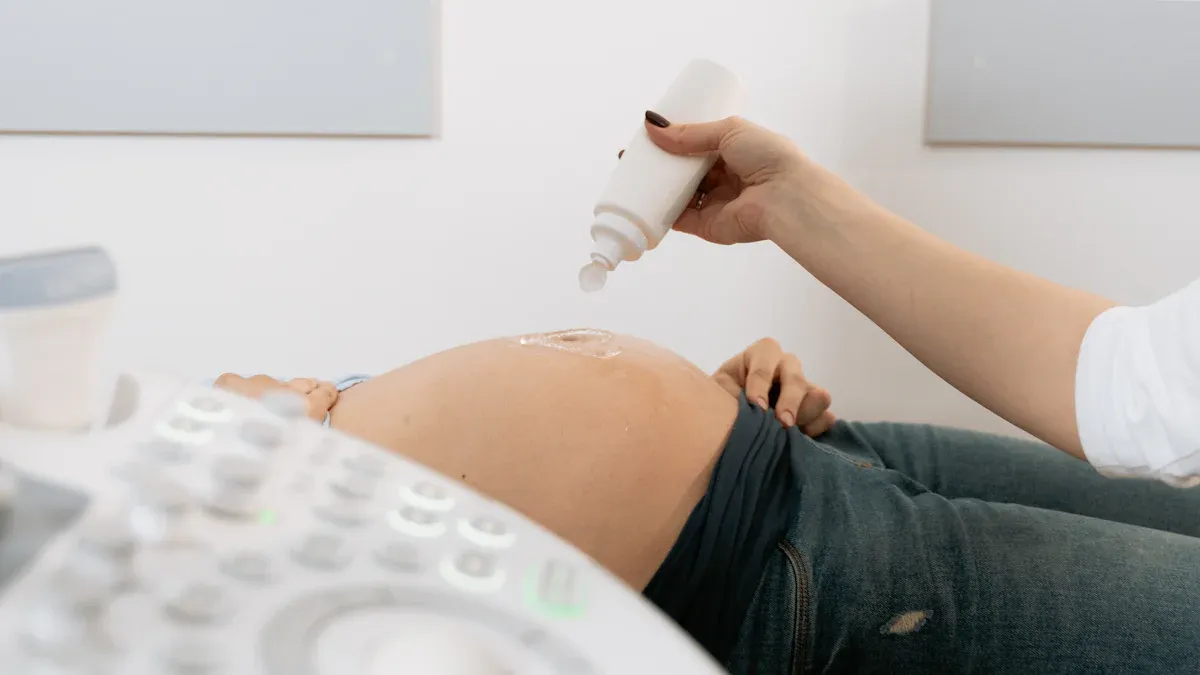
Patients often worry about discomfort or risks during medical imaging. The cartilage detection system uses non-destructive spectroscopic techniques, such as NIR-SWIR absorption and Raman spectroscopy. These methods allow for deep tissue analysis without invasive procedures, ionizing radiation, or the need for contrast agents. Minimal sample preparation makes the process suitable for in situ tissue assessment.
Non-destructive spectroscopic techniques ensure patient safety.
Deep tissue interrogation occurs without invasive procedures.
No ionizing radiation or exogenous contrast agents are required.
Minimal sample preparation supports comfort and convenience.
Patients experience a safer and more comfortable assessment, which encourages participation in regular knee health checks.
Early detection of cartilage degeneration leads to improved long-term outcomes. Machine learning models integrated into the system identify early signs of osteoarthritis with high accuracy. Studies demonstrate that early diagnosis enables timely intervention, which can slow disease progression and enhance quality of life.
| Study | Findings | Accuracy | Method |
|---|---|---|---|
| Mahum et al. | Early KOA detection using ML with high accuracy | 97.14% (cross-validation), 98% (five-fold) | Hybrid feature descriptors (CNN, LBP, HOG) |
| Nagarajan et al. | High detection rate for KOA using PCI | AUC 0.98 | Statistical and geometric feature extraction |
| Kundu et al. | Early detection of OA progression 3 years prior | Test accuracy 0.78 | Cartilage texture maps with transport learning |
Clinicians can intervene earlier, which supports better joint preservation and patient mobility.
Clinicians have observed significant improvements in patient care with the adoption of advanced imaging tools. High-resolution imaging techniques now allow early diagnosis of cartilage injuries. Early detection enables timely interventions, which can improve patient outcomes. Physicians can assess and manage cartilage damage more effectively, leading to better recovery and long-term joint health. Patients benefit from more accurate assessments and personalized treatment plans.
Early diagnosis of cartilage injuries supports timely intervention and improved outcomes for patients.
The cartilage detection system demonstrates measurable gains in accuracy and reliability compared to traditional methods. The following table highlights key performance metrics:
| Measure | Value |
|---|---|
| Smallest Detectable Difference | 0.13 mm |
| Coefficient of Variation (CoV) | 1.3% |
| Mean Point-to-Surface Distance | 0.49 mm |
| Tibia Mean Point-to-Surface Distance | 0.53 mm |
Researchers have confirmed high reliability across diverse patient populations. The system achieves strong intraclass correlation coefficients (ICCs) for various cartilage measurements:
| Measurement Type | ICC Coefficient Range | P-Value |
|---|---|---|
| Global Cartilage | 0.958 to 0.997 | <0.0001 |
| Compartment Measurements | 0.974 to 0.998 | <0.0001 |
| Femoral Condyles | 0.943 to 0.999 | <0.0001 |
| Test-Retest Reliability | 0.978 to 0.999 | <0.0001 |
| Patient Positioning Reliability | 0.978 to 0.999 | <0.0001 |
Clinicians value the system’s quick acquisition speed and cost-effectiveness. The technology fits well into routine clinical workflows and supports both longitudinal and interventional studies.
Researchers continue to push the boundaries of cartilage detection technology. They focus on integrating artificial intelligence with advanced imaging tools. This integration promises to boost the accuracy of cartilage assessments. New developments in 3D convolutional neural networks (CNNs) and vision transformers (ViTs) allow systems to extract more detailed features from medical images. These models improve the system’s ability to understand complex anatomical structures. AI algorithms now analyze large datasets, which helps identify subtle patterns and early abnormalities. This capability supports earlier intervention and better patient outcomes.
The combination of AI and next-generation imaging will likely set new standards for cartilage health evaluation.
Key technological advances include:
Enhanced AI and machine learning integration with imaging platforms
Adoption of 3D CNNs and ViTs for superior feature extraction
Advanced pattern recognition for early detection and intervention
The field of cartilage detection is not limited to knee health. Scientists have started to apply these technologies to other joints. Research now explores cartilage repair in the temporomandibular joint (TMJ) and the hip. Studies in large animal models help validate these approaches for TMJ cartilage repair. The medical community recognizes the need for clear regulatory guidance to support new cartilage repair products for non-knee joints.
Ongoing research targets TMJ and hip cartilage assessment
Recent studies use animal models to refine TMJ repair techniques
Regulatory frameworks will play a role in expanding clinical use
The future of cartilage detection will likely include a broader range of joints, offering hope for patients with diverse joint health needs.
The cartilage detection system transforms knee cartilage checks by using radiomics and machine learning for accessible, early diagnosis. Standardized workflows and predictive analytics support personalized care and efficient resource use. Clinicians and patients benefit from these innovations. Embracing advanced, patient-centered technology will shape the future of joint health.
| Evidence Description | Significance for Patient Care and Clinical Workflow |
|---|---|
| Standardization of medical imaging workflows | Promotes personalized treatments and efficient resource management, leading to cost reductions in health systems. |
| Radiomics systems analyzing patient data | Enables healthcare providers to predict future needs and allocate resources more efficiently. |
The system uses advanced imaging and machine learning. Clinicians receive faster, more accurate results. Patients benefit from early diagnosis and a comfortable experience.
Yes. The system uses non-invasive imaging. It does not require ionizing radiation or contrast agents. Patients experience minimal discomfort during the assessment.
Yes. The system identifies subtle changes in cartilage structure. Early detection supports timely intervention and helps prevent further joint damage.

We provide you with comprehensive foreign trade solutions to help enterprises achieve global development
Automatic recognition and rejection, fish bone ins...
Chicken bone inspection, global poultry AI databas...

Uneven and overlapped product detection, thin & lo...

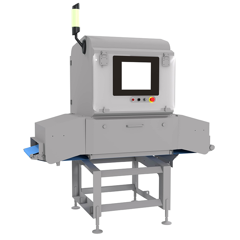
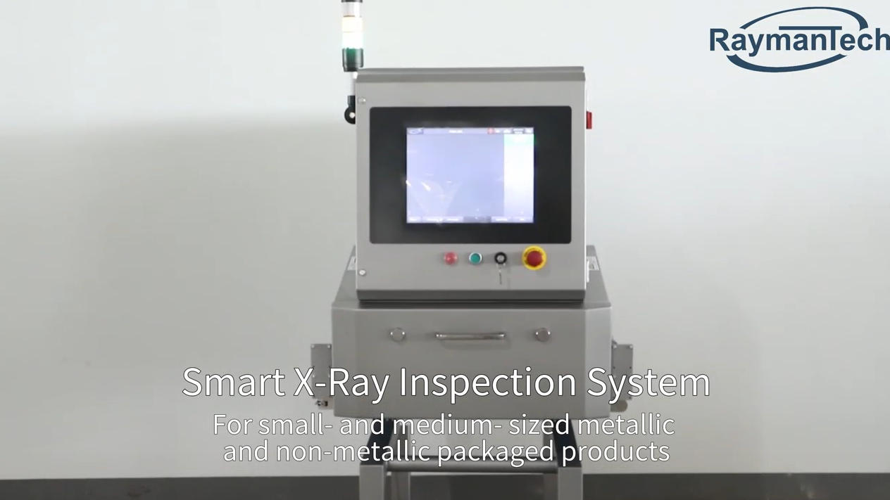
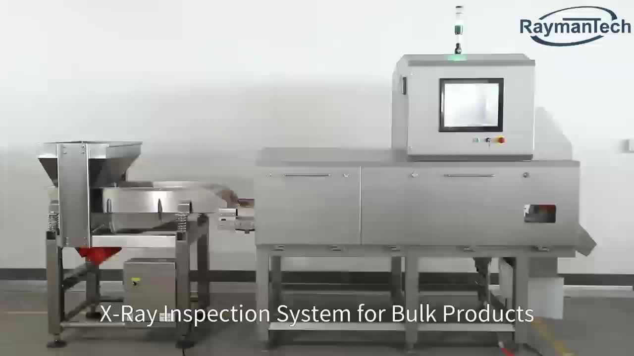
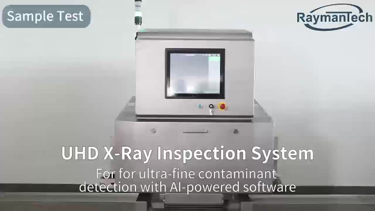
Ultra-fine contaminants detection, UHD X-Ray detec...
Select the most popular foreign trade service products to meet your diverse needs
Select the most popular foreign trade service products to meet your diverse needs


User Comments
Service Experience Sharing from Real Customers
Michael Chen
Orthopedic SurgeonThe Cartilage Detection System has revolutionized our pre-operative planning. Its accuracy in mapping cartilage thickness and lesions is exceptional, allowing for more precise surgical interventions and better patient outcomes.
Sarah Johnson
Senior MRI TechnologistThis system integrates seamlessly with our existing MRI workflows. The automated cartilage segmentation saves us hours of manual contouring, increasing department throughput. The visualization tools are particularly helpful for patient consultations.
David Rodriguez
Sports Medicine ResearcherAs a researcher studying early osteoarthritis, the quantitative data from this system is invaluable. The repeatability and detailed metrics on cartilage volume and health have significantly improved the quality of our longitudinal studies.
Emily Watson
Clinical Applications SpecialistDeploying and training staff on this system was straightforward. The user interface is intuitive, and the support team is responsive. Our clinicians appreciate the clear, actionable reports it generates for both diagnosis and monitoring treatment efficacy.